Radius 3d Anatomy
Anatomy of the Radius
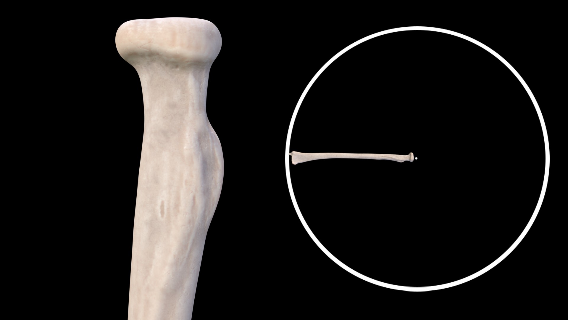
Did you ever wonder why this bone is called the radius?
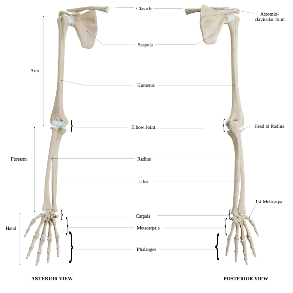
Whenever the forearm pronates or supinates, the Radius rotates relative to the more fixed ulna, which gives the name Radius to this bone. You can see the comparison here in this pic.
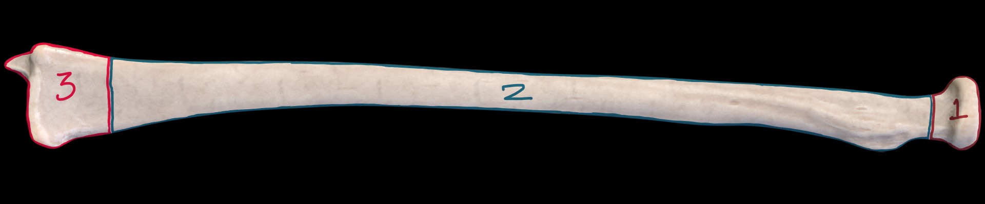
This is the smallest of all the arm bones, namely Radius, Ulna and the Humerus. The Radius ossifies in three centers,
- Proximal Epiphysis
- Shaft
- Distal Epiphysis
Articulations
Proximally, the Radius articulates with the capitulum of the humerus. Distally it articulates with the two carpal bones, the Lunate medially and Scaphoid Laterally. Radius also articulates with the Ulna medially on both the proximal and distal ends.
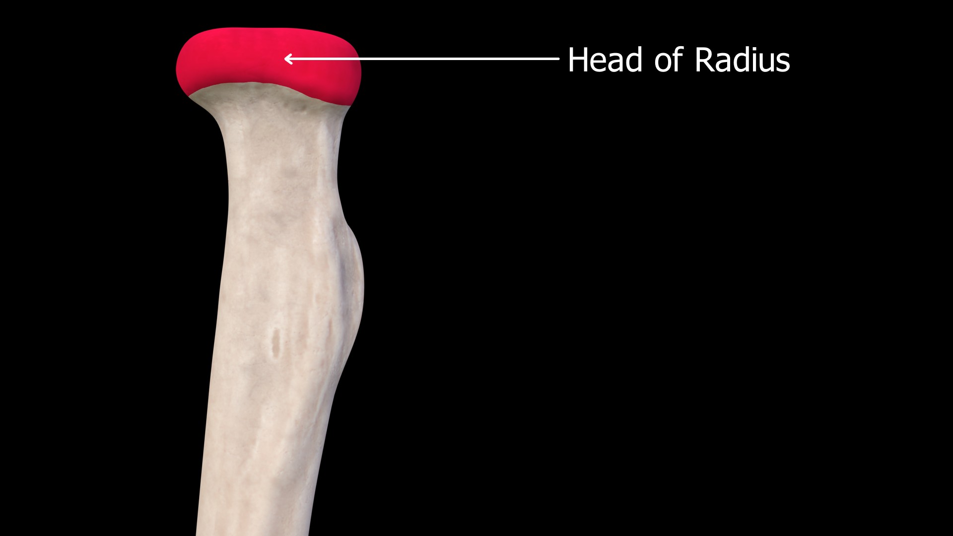
Head of the Radius
The proximal end of the radius has a round articular surface that articulates with the humerus and the ulna.
 This cupped surface is called the Articular facet or Articular fovea. The humerus is attached here.
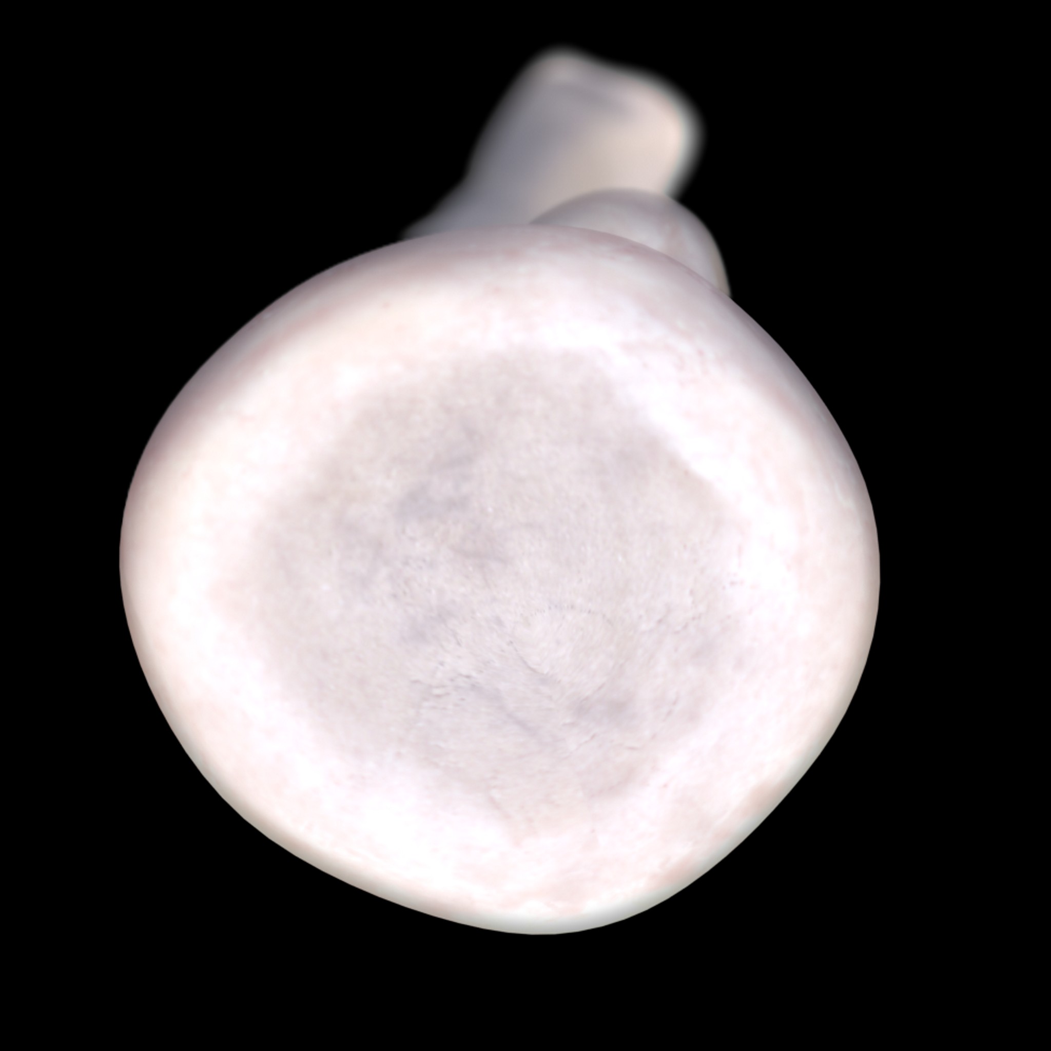 This part is the articular circumference. The Radius is attached medially with the radial notch of ulna here.
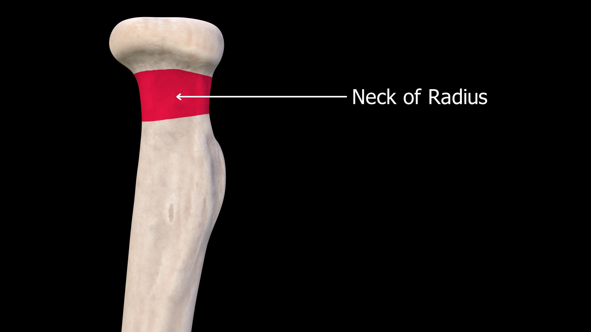
Neck of the Radius
This part between the radial head and the radial tuberosity is termed the Neck of the Radius.
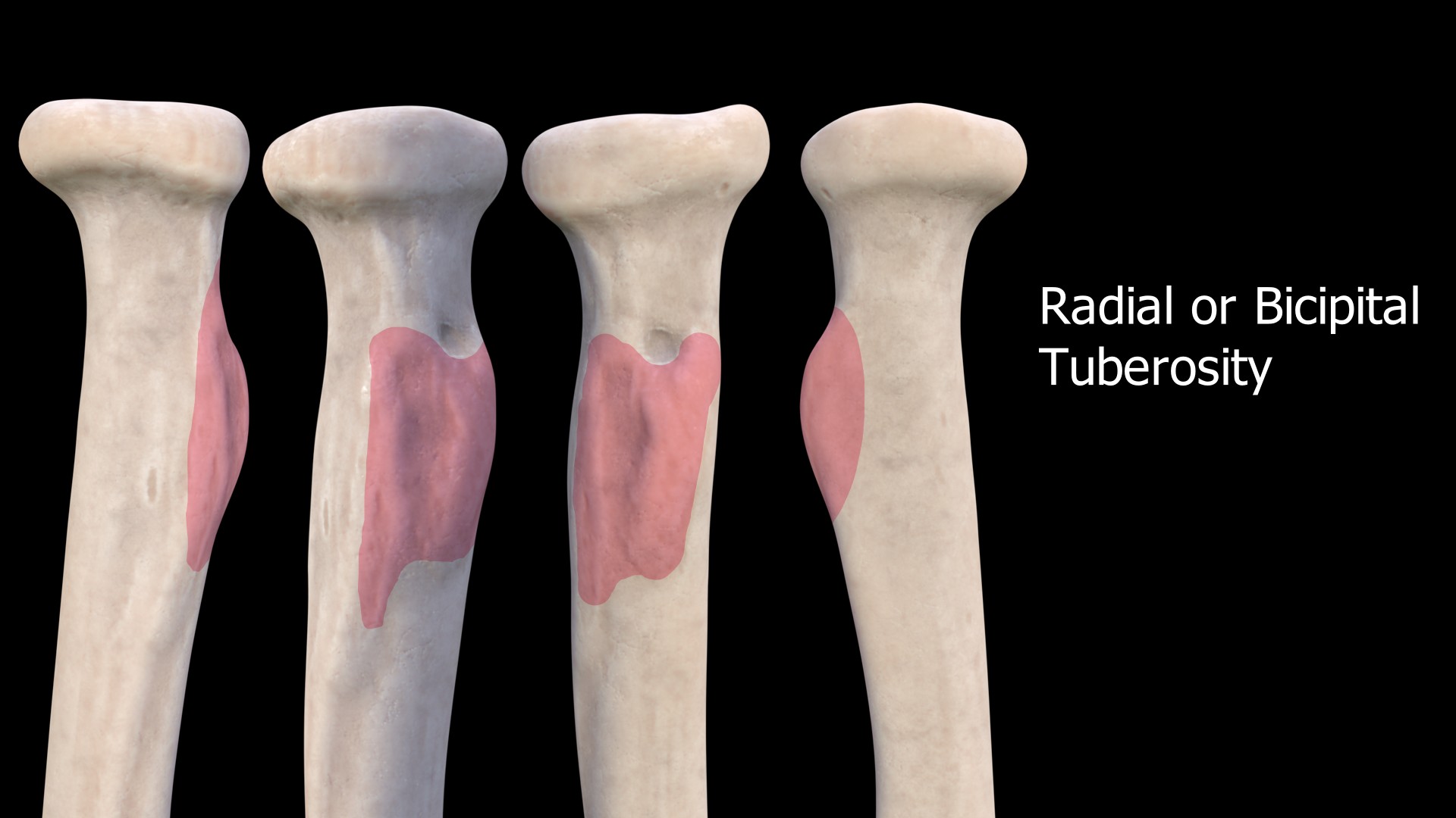
Radial Tuberosity or Bicipital Tuberosity
This blunt protuberance on the anteromedial side is called the Radial Tuberosity or Bicipital Tuberosity. This process is present in the proximal radius and starts after the radial neck. This is where the biceps brachialis is attached.
Shaft of the Radius
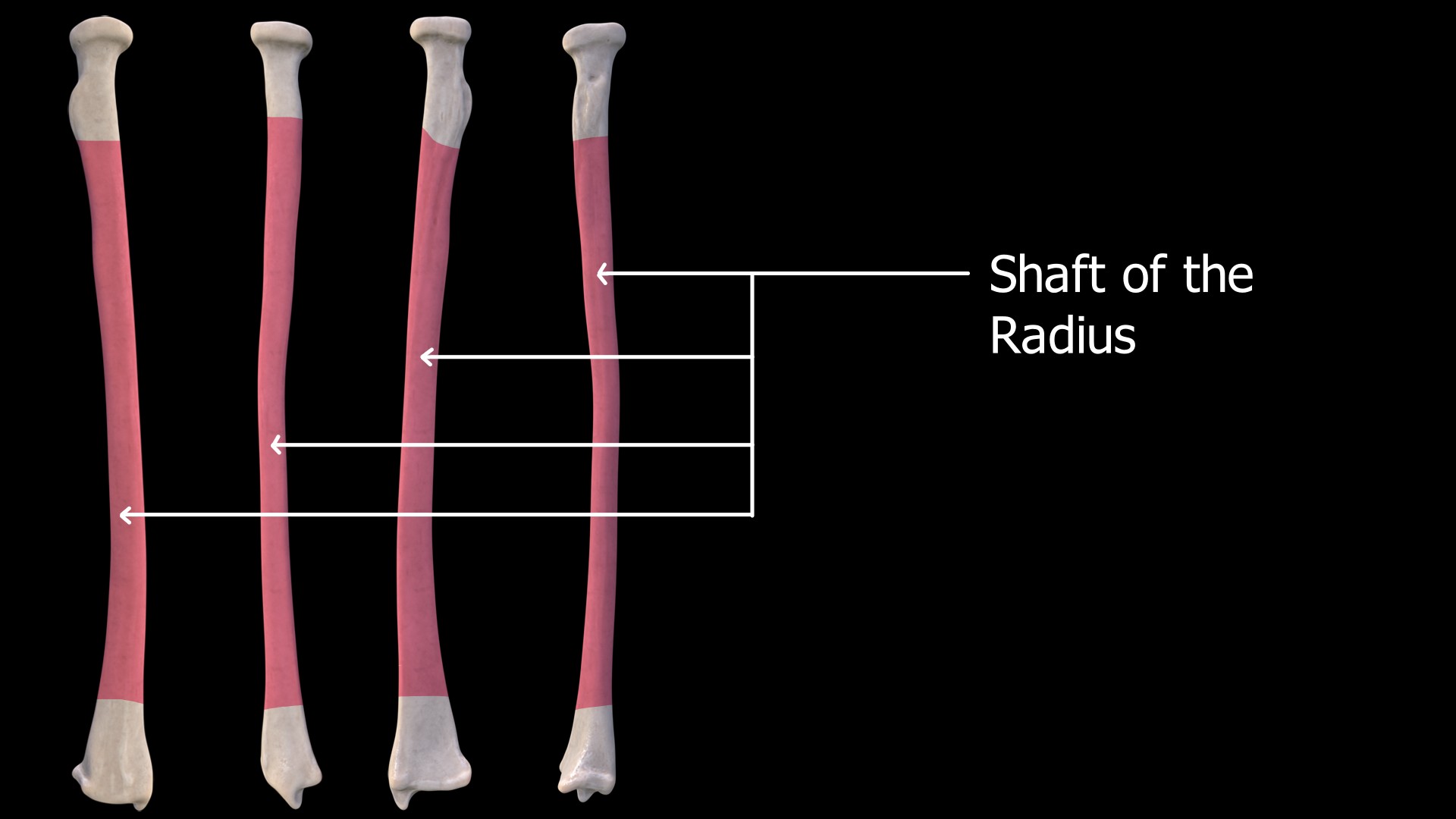 This thin and long segment extending from the radial tuberosity to the distal part of the radius is called the shaft. Three borders divide the shaft into three surfaces.
Borders
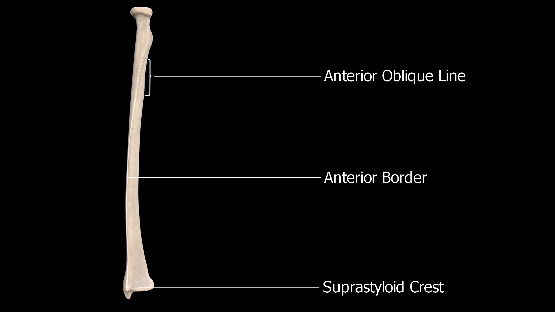
1. Anterior Border
The anterior border extends from the anterior oblique line to the suprastyloid crest.
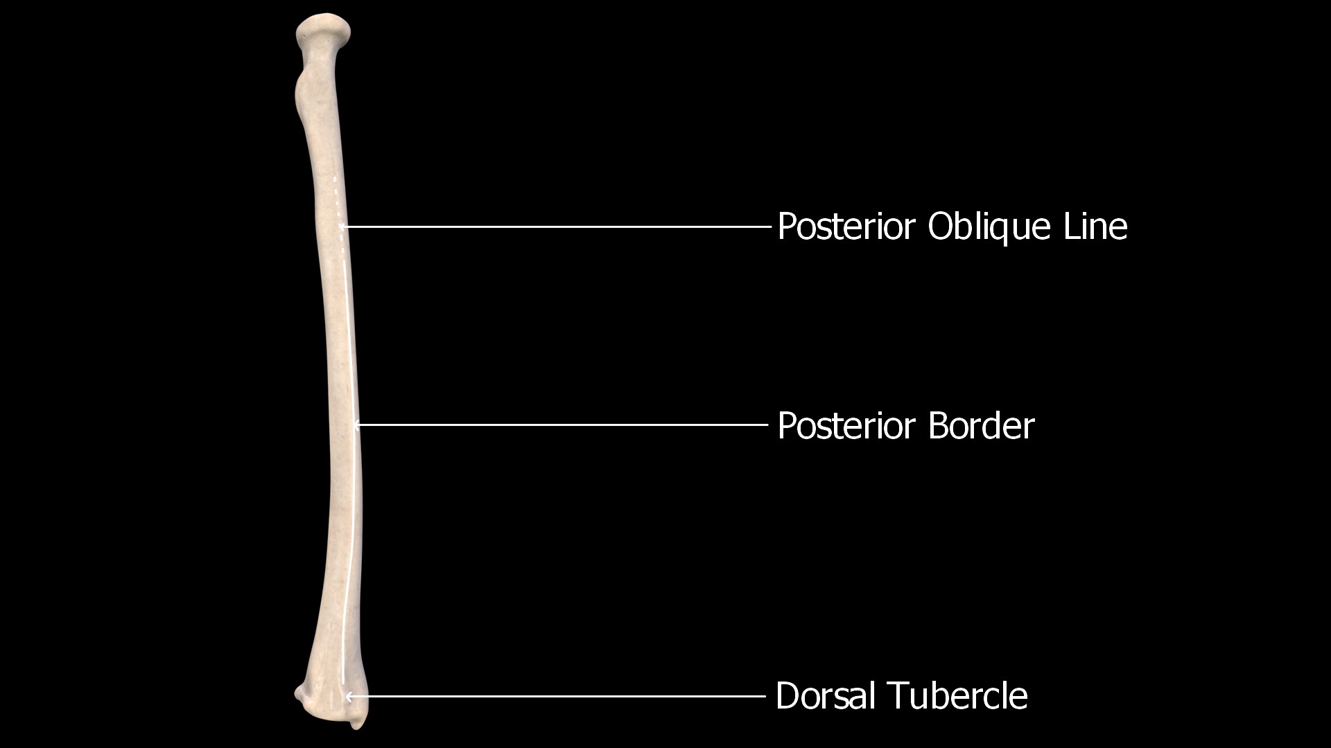
2. Posterior Border
The posterior border is along the posterior aspect and extends from the posterior oblique line to the dorsal tubercle.
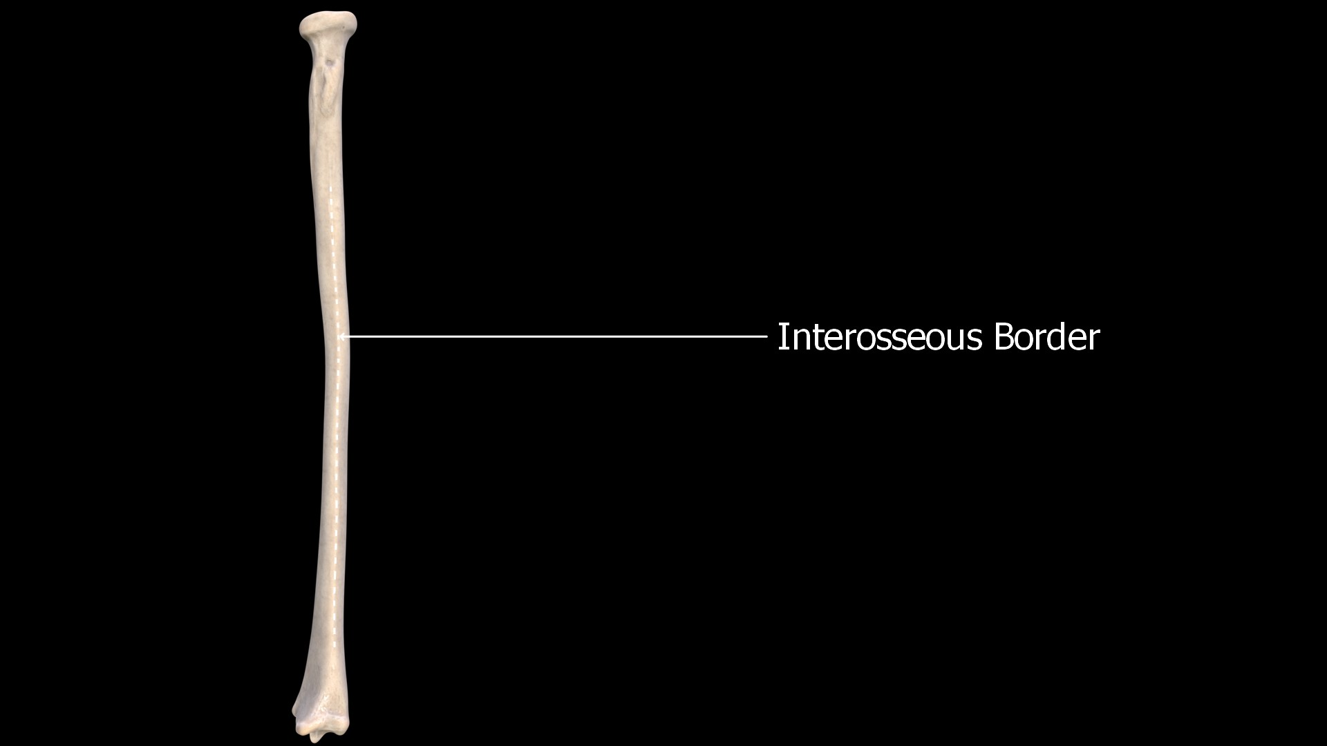
3. Interosseous Border
This sharp edge of the radius is seen on its medial aspect. This is where the interosseous membrane is attached.
Surfaces
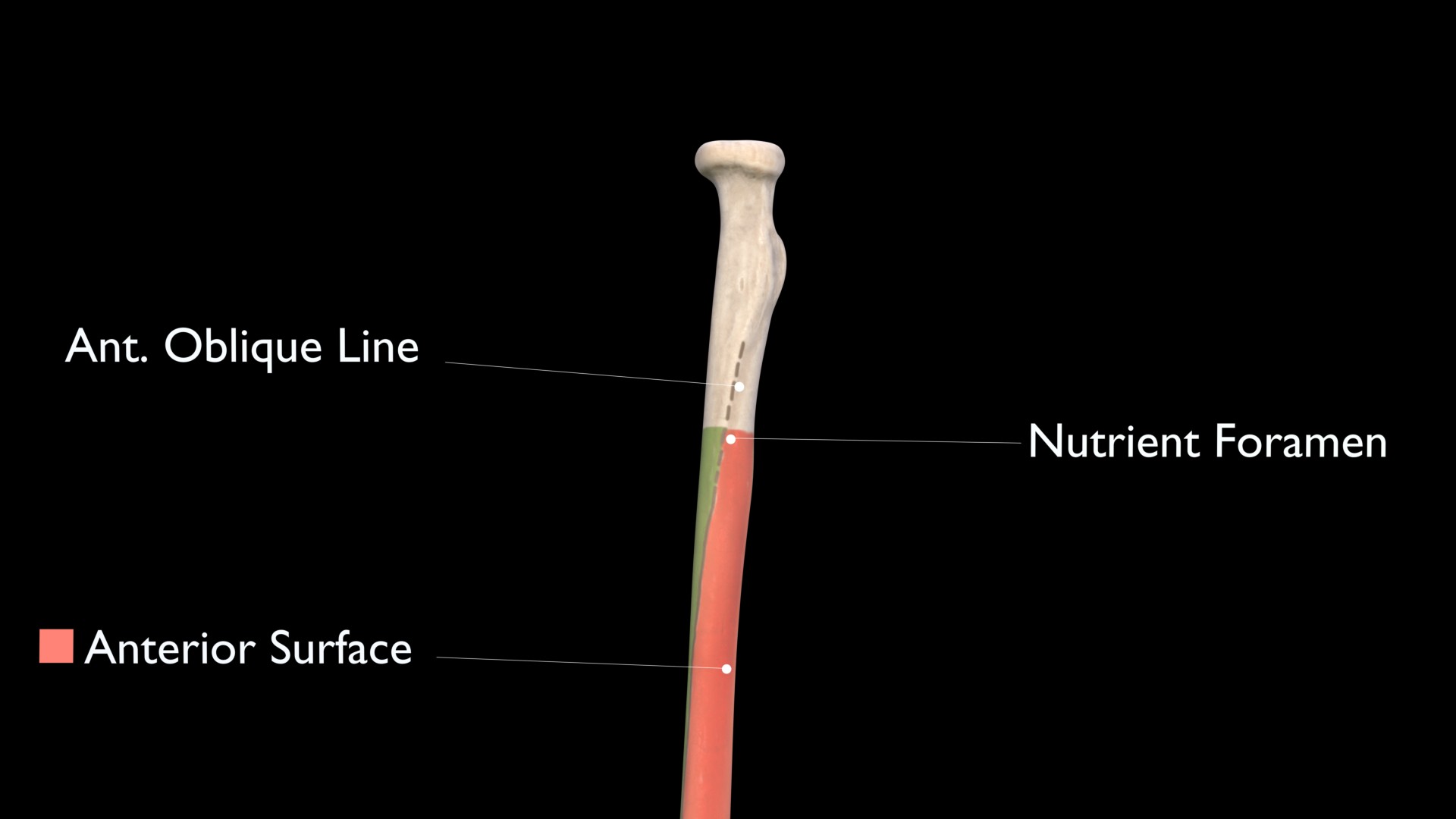
1. Anterior Surface
The anterior surface lies between the anterior and interosseous borders.
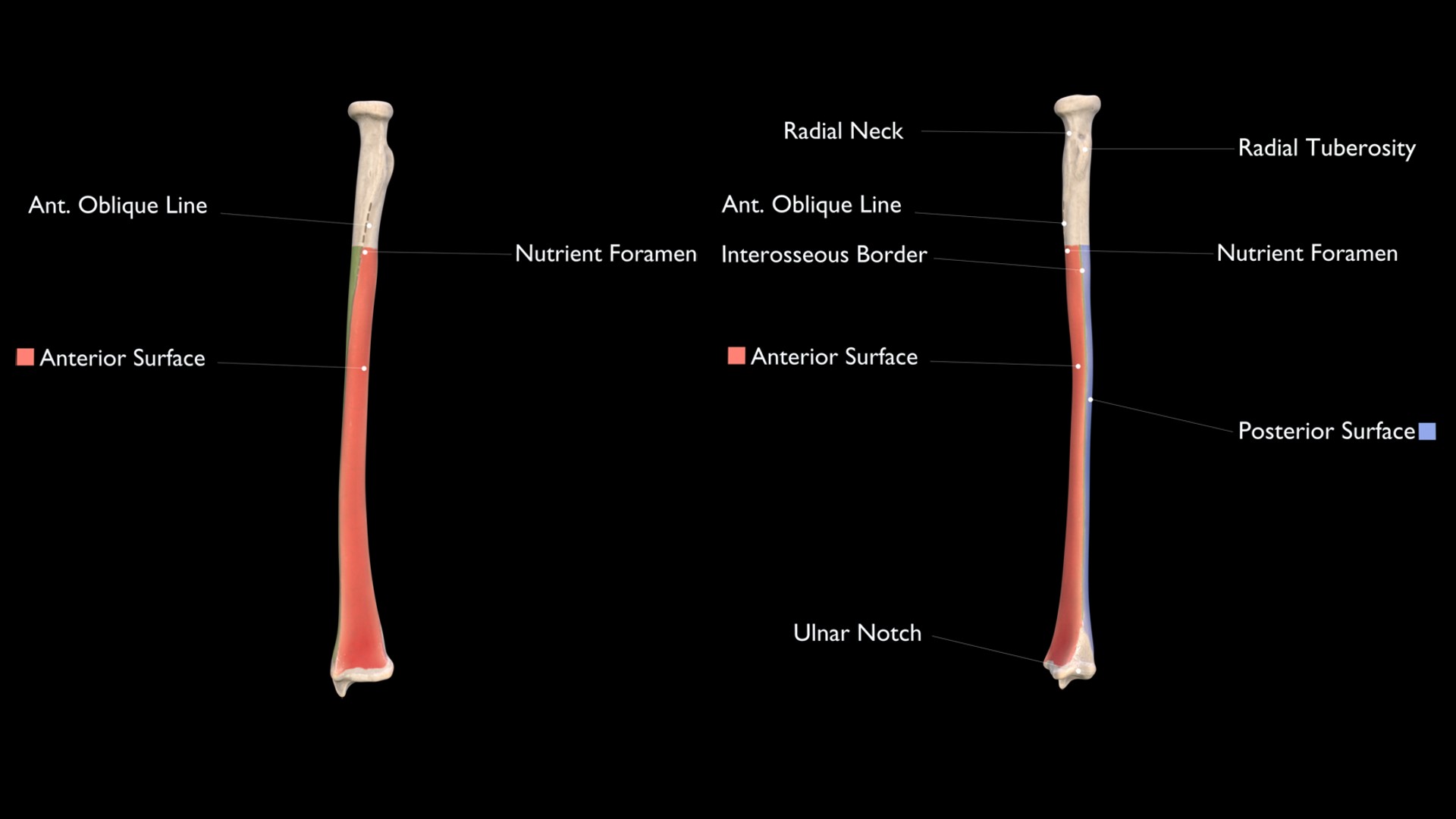
2. Posterior Surface
The posterior surface lies between the posterior and interosseous borders.
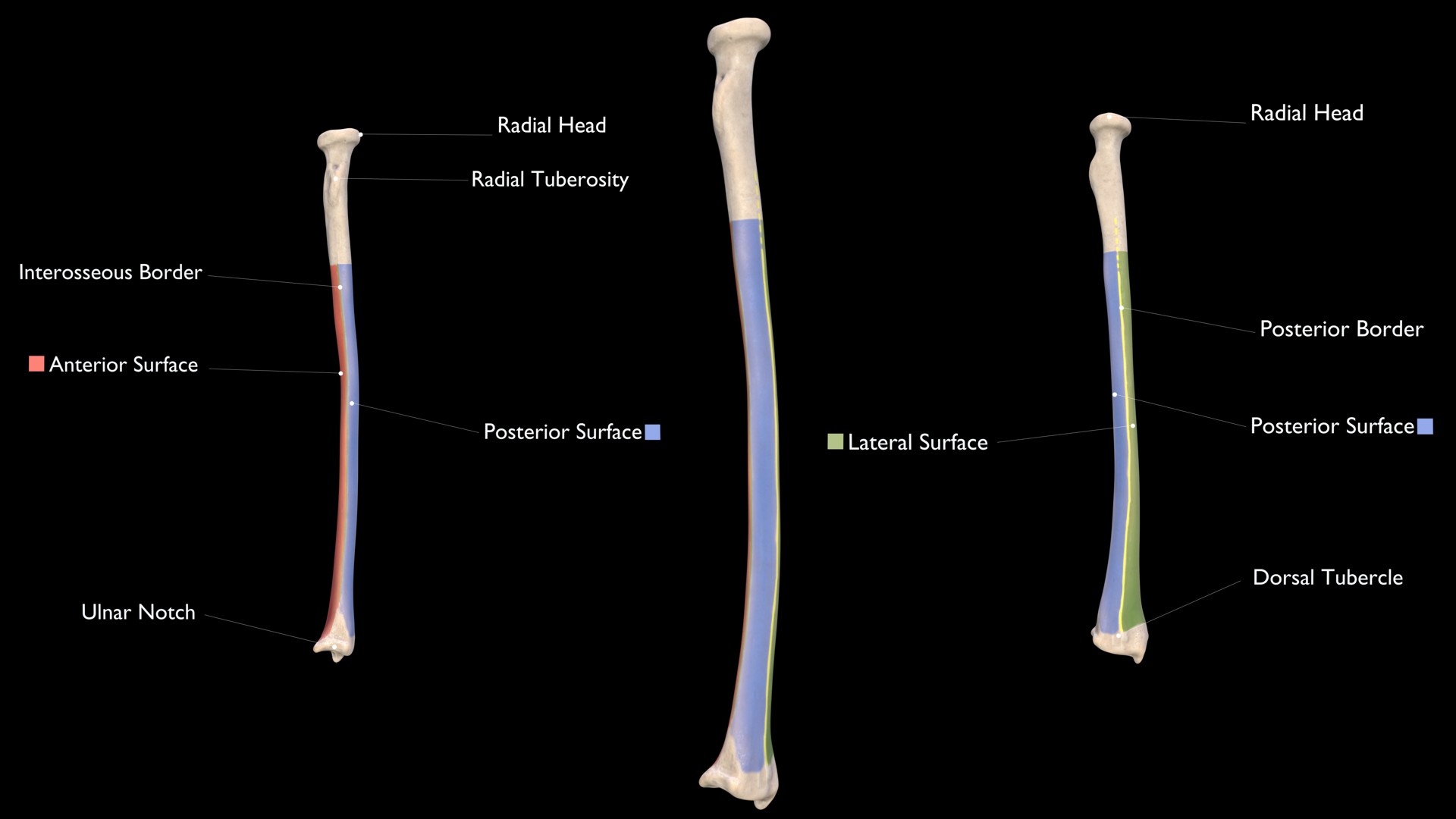
3. Lateral Surface
The lateral surface is the area between the anterior and posterior borders.
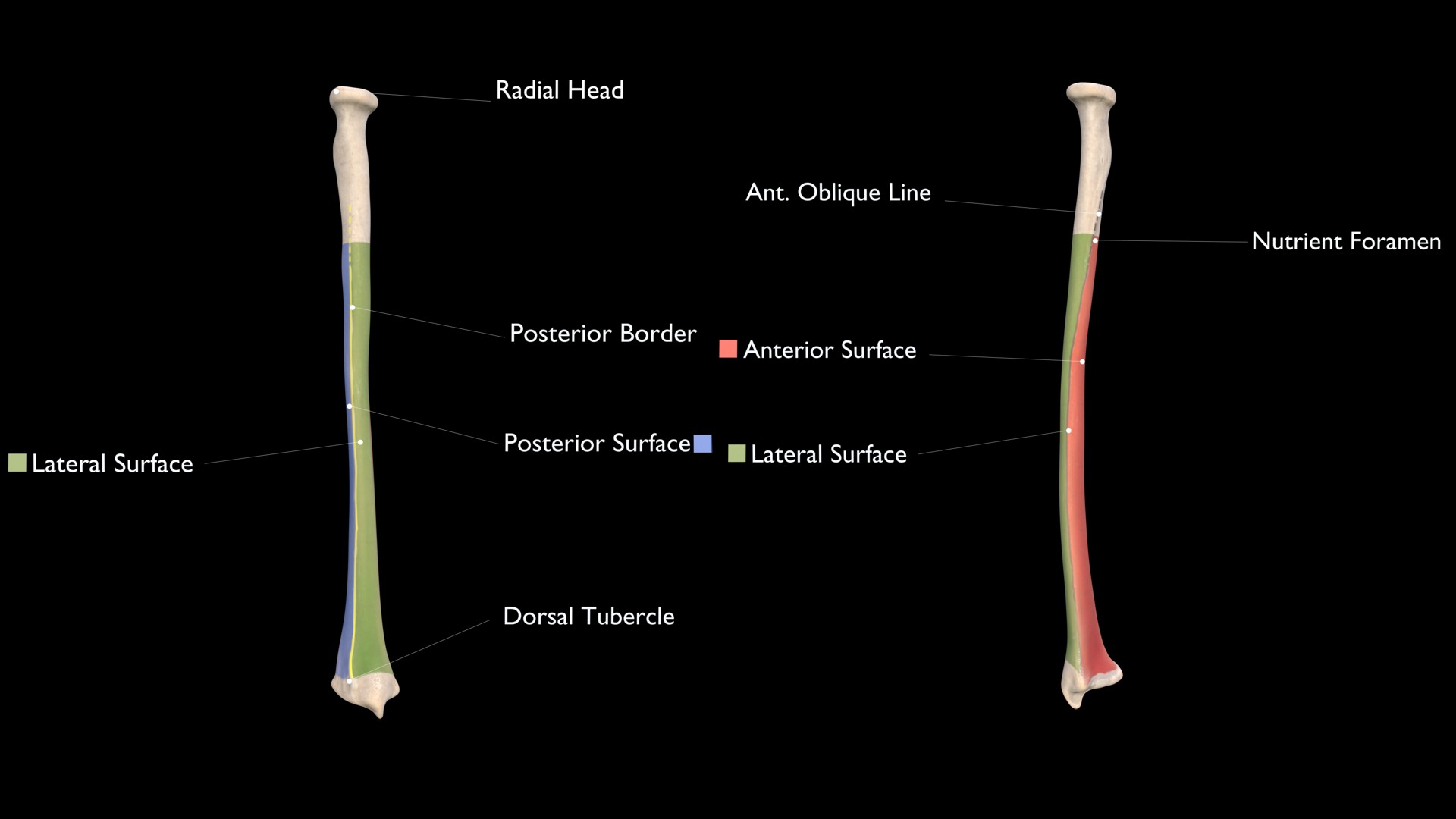
Anterior Oblique Line
The Anterior Oblique line begins from the radial tuberosity anteriorly and spirals inferolaterally and gives origin to the extrinsic muscles of the hand.
Posterior Oblique Line
The Posterior Oblique line begins from the radial tuberosity posteriorly and spirals down to the tuberosity for the pronator teres muscle.
Nutrient Foramen
The nutrient foramen of the Radius is present on the anterior surface of the proximal radius and exits the bone towards its distal end.
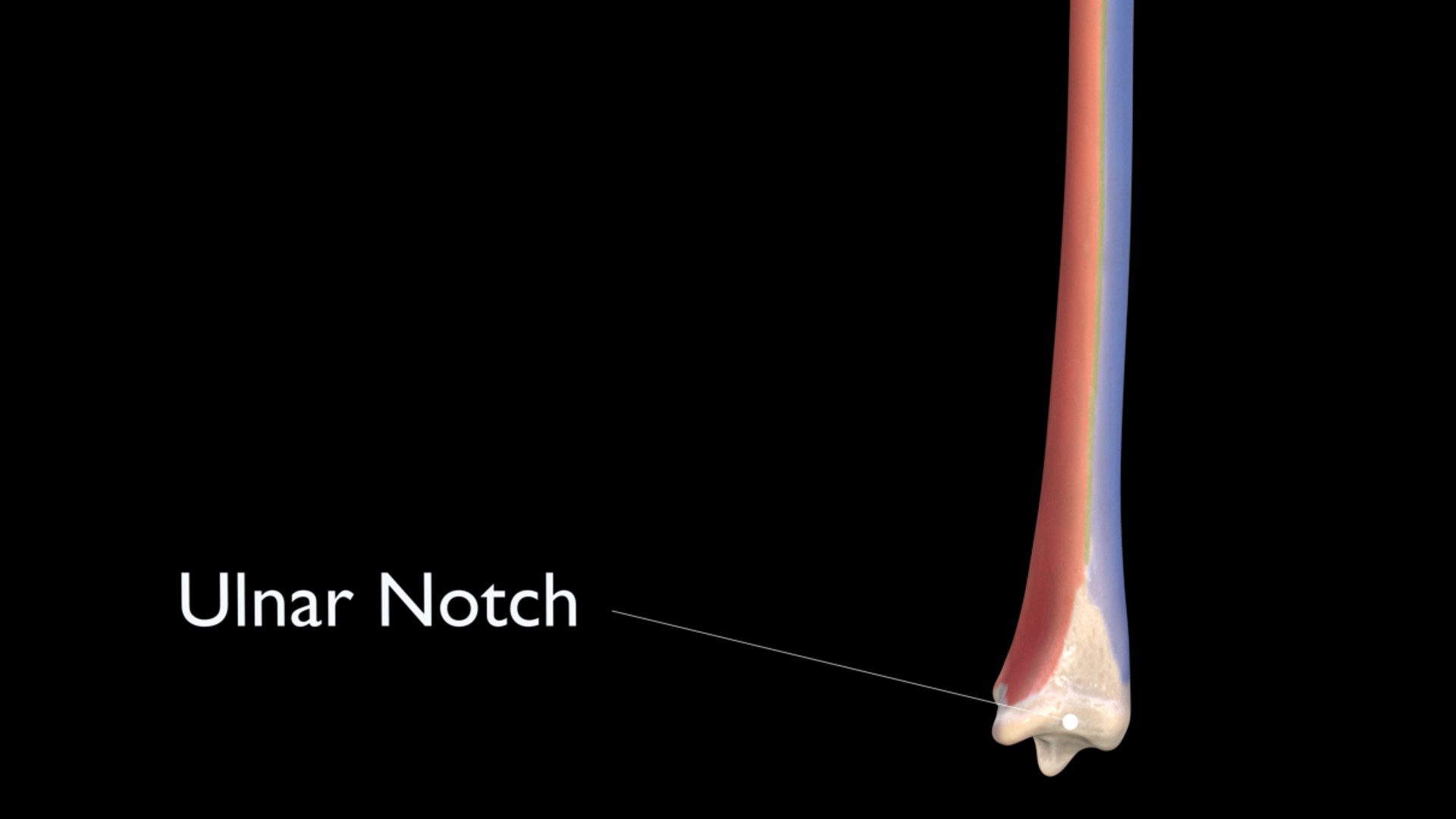
Ulnar Notch
On the distal part of the radius is the articular hollow which is concave in shape. The distal end of the ulna articulates with the distal radius at this concave ulnar notch.
Carpal Articular Surface
The carpal articular surface also termed the distal articular surface of the radius is the distal-most part of the radius, which articulates with the lunate bone on the medial side and the scaphoid bone on the lateral side.
Styloid process
The lateral part of the radius has a sharp projection in its distal part. This is termed the Styloid process or Radial Styloid.
Suprastyloid Crest
The Suprastyloid crest is an oblique crest running from the styloid process. Brachioradialis muscle is attached here.
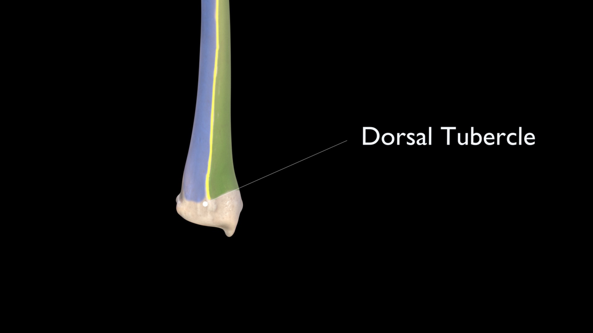
Lister’s tubercle
The distal radius has a large tuberosity called the dorsal tubercle or Lister’s tubercle. It is located on the posterior surface.
Grooves for tendons:
Extensor muscle tendons:
These tendons pass through the posterior surface in the broad depression located on the medial side of the Radius.
Extensor Pollicis Longus:
This tendon passes medial to the Lister’s Tubercle.
Extensor Carpi Radialis:
Passes lateral to the Lister’s Tubercle.

This work is licensed under a Creative Commons Attribution-ShareAlike 4.0 International License.